Search

Light-responsive E. coli functional biofilms as scaffolds for hydroxyapatite mineralization - Phys.org

sekirta.blogspot.com
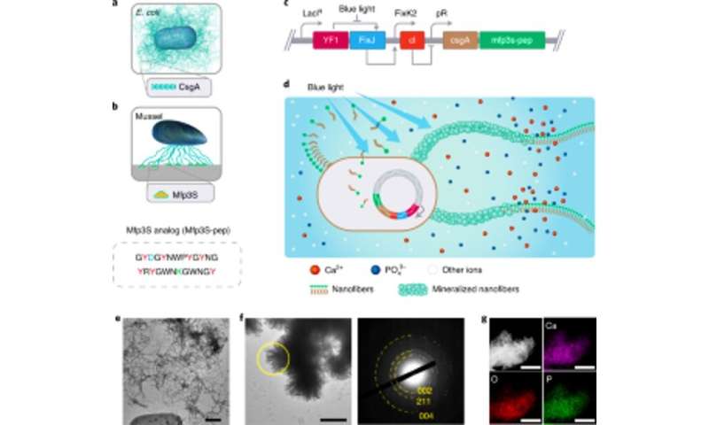
Engineering light-responsive E. coli functional biofilms as scaffolds for HA mineralization.
Engineering light-responsive E. coli functional biofilms as scaffolds for HA mineralization. Engineering light-responsive E. coli functional biofilms as scaffolds for HA mineralization. (a) Schematic illustration of wild-type E. coli biofilms comprising extracellular CsgA amyloid nanofibers, which are major biofilm protein components that contribute to robust adherence of biofilms to solid surfaces. (b) Schematic illustration of Mfps from the adhesive plaques of the California mussel M. californianus. Mfp3S-pep (below) is a bio-inspired adhesive peptide that mimics the original Mfp3S protein found in the adhesive plaques of M. californianus. Mfp3S-pep is enriched with aspartic acid (blue), lysine (green) and tyrosine (red) residues, known for their functional roles in promoting the nucleation, growth and adhesion of HA. (c) Schematic showing the genetic components of the light-responsive E. coli biofilm. The design of functional biofilms was enabled by fusing sequences encoding the CsgA protein and Mfp3S-pep, both positioned downstream of the light-sensitive pDawn transcriptional control element. In the pDawn circuit, the constitutive expression of histidine kinase YF1 and its cognate response regulator FixJ is tightly regulated by the LacIq promoter, while expression of the λ phage repressor cI is controlled by the FixK2 promoter. Upon blue-light illumination, kinase activity of YF1 and consequent expression of cI are both inhibited, which in turn activates the λ promoter pR to promote the expression of CsgA–Mfp3S-pep. (d) Schematic showing local deposition of HA minerals on the functional extracellular nanofibers composed of CsgA–Mfp3S-pep fusion proteins, secreted by engineered light-responsive E. coli, the lightreceiver-CsgA– Mfp3S-pep strain. (e) TEM image showing cells and extracellular matrix with abundant amyloid fibers in the biofilms. Scale bar, 500 nm. f, TEM image showing the formed composite and lath-like crystals precipitated on the surfaces of extracellular nanofibers after 7 d of mineralization in 1.5× SBF. The corresponding SAED pattern presents diffraction arcs assigned to the (002), (211) and (004) planes. Scale bar, 500 nm. g, EDS mapping illustrating the Ca, O and P elements intrinsically assigned to the HA phase. Scale bars, 500 nm. Credit: Nature Chemical Biology, doi:https://ift.tt/3hI0Jnb

Living organisms have evolved mechanisms of biomineralization to build structurally ordered and environmentally adaptive composite materials. While research teams have significantly improved biomimetic mineralization research in the lab, it is still difficult to engineer mineralized composites with structural features and living components much like their native counterparts. In a new report now published on Nature Chemical Biology, Yanyi Wang and a research team in physics, advanced materials, synthetic biology, and engineering in China, developed living patterned and gradient composites inspired by natural graded materials. They coupled light-inducible bacterial biofilm formation with biomimetic hydroxyapatite (HA) mineralization in this work, to show how the location and degree of mineralization could be controlled. The cells in the composites remained viable while sensing and responding to environmental signals. The composites showed a 15-fold increase in Young's modulus (i.e., stiffness, the ratio between stress and strain) after mineralization. The work sheds light to develop living composites with dynamic responsiveness and environmental adaptability.

Biomineralization in the lab

Living organism can produce a variety of hierarchical organic-inorganic composite structures based on biomineralization, where notable examples include the radular teeth of chitons, fish scales and crayfish mandibles that fulfill diverse biological functions. The timing and degree of biomineralization must be precisely controlled by cells to form accurate structures and functions in living organisms. Recent research in biomimetic mineralization had highlighted the importance of exploring cell-controlled approaches to produce living patterns and graded composites as a promising yet largely unexplored area. The disciplines of synthetic biology and materials science have unleashed a range of sophisticated and environmentally friendly gene circuits to bioengineer a range of new cellular functions. In this work, Wang et al. developed bioinspired, living composite materials by harnessing light-inducible Escherichia coli biofilms coupled with biomimetic mineralization. The work will open doors to integrate engineered cells to produce mineralized materials with structural and living features.
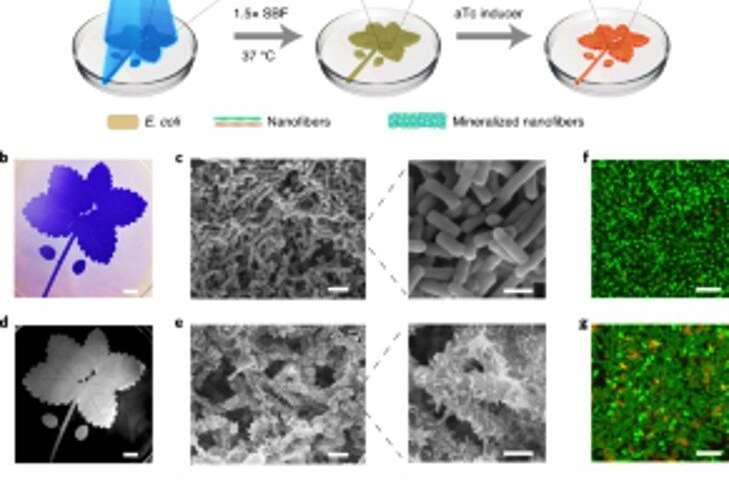
Engineering light-responsive E. coli functional biofilms as scaffolds for HA mineralization.
Spatially controllable mineralization of light-inducible biofilms for living patterned composites. (a) Schematic of the experimental setup and sequential steps for producing living patterned composites. The blue leaf pattern depicted in the illustration was projected onto a Petri dish containing the lightreceiver-CsgA–Mfp3S-pep strain suspended in medium. Blue light triggers functional biofilm formation through the light-regulated expression of CsgA–Mfp3S-pep proteins from the lightreceiver-CsgA–Mfp3S-pep strain. The culture medium in the Petri dish was then replaced with 1.5× SBF, followed by incubating at 37 °C to form composites. After the mineralization process, aTc was added to induce the expression of a red fluorescent protein (mCherry). (b) Digital camera image of the patterned biofilms stained with CV. Scale bar, 1 cm. (c) SEM images showing the surface morphology of the patterned biofilms. Scale bars: 2 µm (left), 1 µm (right). (d) Digital camera image of the subsequent mineralized composite that retained the original pattern. Scale bar, 1 cm. (e) SEM images showing the surface microstructures of the mineralized composite. Scale bars: 2 µm (left), 1 µm (right). (f,g) Confocal laser scanning microscopy analysis of bacterial viability in biofilms (f) and composites (g). Scale bars, 5 µm. Note that SYTO 9 dye and PI were applied as labeling agents to stain live (green) and dead (red) cells, respectively. (h) Induced fluorescence of the living composite recorded with a ChemiDoc XRS system. Scale bar, 1 cm. i, Confocal microscopy demonstrated mCherry expression from the bacteria in the composite. Scale bar, 20 µm. j, Confocal images of a living composite after inducing mCherry expression and labeling with calcein, which indicated a homogeneous distribution of organic components and inorganic minerals inside the patterned living composites. Scale bars, 20 µm. Credit: Nature Chemical Biology, doi:https://ift.tt/3hI0Jnb
Selecting protein modules for hydroxyapatite (HA) mineralization and developing a light-sensitive biofilm

The team selected fusion proteins to engineer mineralization promoting E. coli biofilms. Based on previous experiments, they selected protein Mefp5 – originating from Mytilus edulis, followed by Mfp3S – originating from Mytilus californianus and another variant of the Mfp3S peptide (Mfsp3S-pep) to initiate mineralization and promote adhesion. The team constructed fusion proteins containing a major protein domain of the E. coli to form CsgA-Mfp fusion proteins and confirmed their potential secretion from engineered cells. They then selected the CsgA–Mfp3S-pep fusion protein as a representative for hydroxyapatite mineralization and conducted experiments to verify the function of the protein to highlight their role during mineralization and crystal formation. Thereafter, Wang et al. constructed a light-inducible biofilm-forming strain named lightreceiver-CsgA-Mfp3S-pep that can be tightly regulated via blue light illumination.

The light-sensitive strain could generate functional biofilm materials after illumination with light to promote the mineralization of hydroxyapatite (HA). To validate this, the scientists exposed the light-sensitive strain to blue light in a Petri dish and used histological staining and transmission electron microscopy (TEM) imaging to show the production of amyloid fibers in the biofilms. Comparatively, they did not observe amyloid fibers in samples grown in the dark. The engineered extracellular matrix also acted as a template for HA mineralization in time, which they confirmed after 7-days of incubation based on X-ray diffraction (XRD) and energy-dispersive X-ray spectroscopy (EDS) techniques.
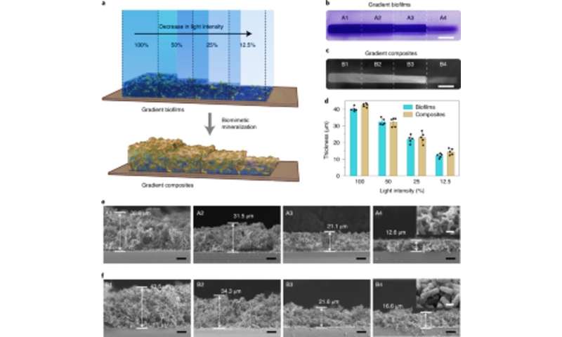
Engineering light-responsive E. coli functional biofilms as scaffolds for HA mineralization.
Density-controllable mineralization in light intensity-regulated gradient biofilms used to fabricate living graded composites. (a) Schematic illustration showing the generation of living gradient composites through in situ mineralization of biofilms with gradient biomass densities. The gradient biofilms were formed by projecting light with varying intensities and precise spatial control onto the Petri dish containing the lightreceiver-CsgA–Mfp3S-pep strain suspended in medium. (b) Digital camera image showing CV-stained biofilms with gradient biomass densities. Scale bar, 1 cm. (c) Digital camera image of subsequently formed gradient living composites after HA mineralization. Scale bar, 1 cm. (d) The thickness of biofilms and the subsequently mineralized composites formed under different light intensities. Results are presented as mean ± s.d. Data are representative of n= 5 independent experiments. (e) Sectional SEM micrographs showing the morphologies of different regions (A1–A4) in the light intensity-graded biofilms. Scale bars, 10 µm. The inset image (taken from the A4 region) represents the typical surface morphology of biofilms. Scale bar, 1 µm. (f) Sectional SEM micrographs showing the morphologies of different regions (B1–B4) in the living gradient composites as templated by light intensity-graded biofilm scaffolds. Scale bars, 10 µm. The inset image (taken from the B4 region) represents the typical surface morphology of the mineralized gradient composites. Scale bar, 1 µm. Note that the opacity percentages of 100%, 50%, 25% and 12.5% correspond to actual light intensities of 0.0306 W cm−2, 0.0268 W cm−2, 0.0178 W cm−2 and 0.0140 W cm−2, respectively. Credit: Nature Chemical Biology, doi:10.1038/s41589-020-00697-z
Forming controlled living composites

Based on the light-sensitive nature of the engineered biofilms, Wang et al. manipulated biofilm formation in space-time to control the formation of composites. They accomplished this by illuminating a leaf pattern on liquid bacterial cultures in polystyrene petri dishes that faithfully recapitulated the projected light pattern on to the bacterial biofilms. After 7-days of mineralization, the composite generated in the petri dishes retained the original pattern as observed using scanning electron microscopy. The light-regulated approach controlled the shape of the composite based on grid-pattern projections and spatial resolution of light in the microscale—comparable to the size of the living bacteria. The team then verified the viability of live, intact cells by engineering the living composites to express fluorescent proteins, as confirmed using confocal microscopy images. Thereafter, they used thermogravimetric analysis to quantify the inorganic components of mineralized composites, where the inorganic material increased proportionally with time on immersion in simulated body fluid (SBF). Wang et al. also compared the Young's modulus of the biofilm using a micro-indentation technique to show how mineralization strengthened the E. coli biofilms to protect the cells.

Density controlled gradient composites
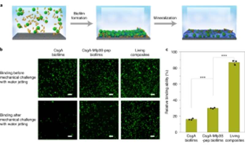
Engineering light-responsive E. coli functional biofilms as scaffolds for HA mineralization.
Coupling engineered biofilms with mineralization for robust capturing and immobilization of microspheres on substrates. (a) Schematic showing the capture and immobilization of microspheres on a glass slide in solution. PS microspheres suspended in culture medium were captured and immobilized on a substrate through biofilm formation followed by mineralization in 1.5× simulated body fluid (SBF). (b) Fluorescent images showing the two types of biofilms (left and center columns) and living composite-immobilized microspheres (right column) on the substrates before (top) and after (bottom) challenge with water jetting at a constant discharge pressure of 8 psi. Scale bars, 100 µm. (c) Quantification of the relative capabilities of different biofilms and living composites to glue and immobilize PS microspheres on the substrate. Results are presented as mean ± s.d. From left to right: P= 0.00003, P= 0.000004. *P 0.05, **P 0.01, ***P 0.001. Statistics are derived using a two-sided t-test. Data are representative of n= 3 independent experiments. Credit: Nature Chemical Biology, doi:https://ift.tt/3hI0Jnb

Tissue engineering approaches have emphasized the need to precisely identify between hard and soft tissues for successful physiological performance, therefore the light intensity could be tuned to control the density and mechanical properties of engineered living materials. Zhang et al. accomplished this by exposing E. coli cultures to different intensities of illumination to reveal how biofilm thickness decreased with the decreasing intensity of blue light. They further showed how mineralization was tightly and locally directed by the engineered Mfsp3S-pep fusion proteins. Then they studied the local mechanical properties of illuminated regions with micro-indentation to observe a fourfold increase in Young's modulus to highlight living composites engineered with tunable mineral gradients and mechanical properties for regenerative tissue-to-bone interfaces in bone tissue engineering applications.

Deploying living composites for site-specific damage repair

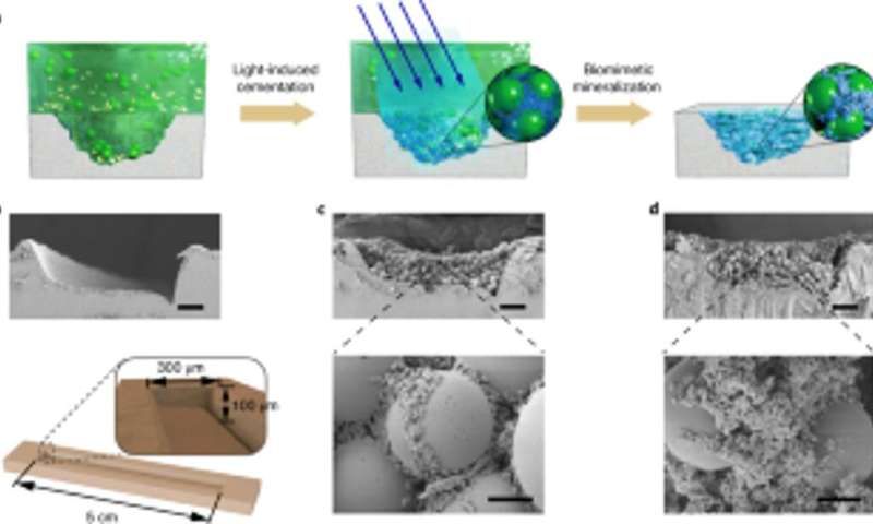
The scientists also studied the capacity for the engineered biofilms to aggregate for damage repair by gluing polystyrene microspheres, alongside HA-mineralization. As proof-of-concept, they applied living mineralized composites to fill and repair cracks engineered on the surface of a polystyrene Petri dish. During the experiments, the light-induced adhesive biofilms captured microspheres in solution to fill the purposely created damaged furrow, while nanofibers in biofilms acted as a template for hydroxyapatite mineralization to consolidate light-induced cementation under blue light illumination. Using scanning electron microscopy, Wang et al. showed how bacteria and the surrounding extracellular matrix adhered together with microspheres to illustrate the adhesive function of the living biofilms. The HA-mineralized composites formed dense, concrete-like layers that filed the damaged furrow to highlight the mineralization functionality of the living composites for enhanced durability and repair applications.
Engineering light-responsive E. coli functional biofilms as scaffolds for HA mineralization.
Application of controllable living mineralization for site-specific damage repairs. (a) Schematic showing application of living mineralized composites to fill and repair cracks on the surface of a PS Petri dish. From left to right: damage furrow on the Petri dish substrate, light-induced adhesive biofilms capture microspheres in solution to fill the damage furrow, and nanofibers in biofilms act as a template for HA mineralization to consolidate the cement. (b) SEM image showing the damage furrow. Below, a schematic diagram illustrating the dimensions of the damage furrow. Scale bar, 50 µm. c, SEM images showing the morphology of the damage furrow filled with the biofilm and microspheres. Scale bars: 50 µm (top), 5 µm (bottom). (d) SEM images showing the morphology of the damage furrow after mineralization; HA fills in the gaps between spheres. Scale bars: 50 µm (top), 5 µm (bottom). Note that the site-specific damage-repairing experiments were repeated at least three times with identical results. Credit: Nature Chemical Biology, doi:10.1038/s41589-020-00697-z
Outlook

In this way, Yanyi Wang and colleagues produced bioinspired living patterned composites and living graded composites with a light-regulated biofilm platform. The resulting composites displayed environmental responsiveness similar to their native counterparts. The cells within the composites remained viable and responded to environmental stimuli even after mineralization. The work opens a path to create living, functional organic-inorganic composites with adaptive and self-repairing features with applications across biomedicine, regenerative medicine, and bioremediation.


Explore further

Research identifies nanoscale effect of water and mineral content on bone

More information: Wang Y. et al. Living materials fabricated via gradient mineralization of light-inducible biofilms, Nature Chemical Biology, doi: doi.org/10.1038/s41589-020-00697-z

Kröger N. et al. Self-assembly of highly phosphorylated silaffins and their function in biosilica morphogenesis, Science, 10.1126/science.1076221.

Davis S. A. et al. Bacterial templating of ordered macrostructures in silica and silica-surfactant mesophases. Nature, doi.org/10.1038/385420a0

© 2020 Science X Network

Citation: Light-responsive E. coli functional biofilms as scaffolds for hydroxyapatite mineralization (2020, December 31) retrieved 31 December 2020 from https://ift.tt/3hvLVHQ

This document is subject to copyright. Apart from any fair dealing for the purpose of private study or research, no part may be reproduced without the written permission. The content is provided for information purposes only.

Let's block ads! (Why?)


"light" - Google News
December 31, 2020 at 06:00PM
https://ift.tt/3hvLVHQ

Light-responsive E. coli functional biofilms as scaffolds for hydroxyapatite mineralization - Phys.org
"light" - Google News
https://ift.tt/2Wm8QLw
https://ift.tt/2Stbv5k

Bagikan Berita Ini

0 Response to "Light-responsive E. coli functional biofilms as scaffolds for hydroxyapatite mineralization - Phys.org"

Post a Comment

Powered by Blogger.